|
|
||||||||||||
| HOME I ABOUT DR. I SERVICES I FAQ I CONTACT ME I ENQUIRY |
| SERVICES |
| Head & Neck Cancer |
For more information, contact the National Cancer Helpline 1800 200 700 to speak to one of our specialist cancer nurses. DiagnosingFirst, visit your family doctor (GP) or dentist if you are worried about any symptoms. They can examine you and do some blood tests. If your GP or dentist is still concerned about you, they can refer you to a hospital for more tests. You may be referred to a specialist doctor, such as a maxillofacial surgeon or an ENT (ear, nose and throat) specialist. The specialist will discuss your symptoms and examine you again. He or she will inspect your mouth, throat, tongue, nose and neck using a small mirror and/or lights. Your neck, lips, gums and cheeks will also be checked for lumps. The following tests can diagnose mouth, head and neck cancers.
|
| Lung Cancer |
| What Is Lung Cancer? |
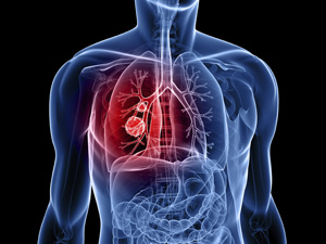
 Lung cancer is the uncontrolled growth of abnormal cells that start off in one or both lungs; usually in the cells that line the air passages. The abnormal cells do not develop into healthy lung tissue, they divide rapidly and form tumors. As tumors become larger and more numerous, they undermine the lung’s ability to provide the bloodstream with oxygen. Tumors that remain in one place and do not appear to spread are known as “benign tumors”. Lung cancer is the uncontrolled growth of abnormal cells that start off in one or both lungs; usually in the cells that line the air passages. The abnormal cells do not develop into healthy lung tissue, they divide rapidly and form tumors. As tumors become larger and more numerous, they undermine the lung’s ability to provide the bloodstream with oxygen. Tumors that remain in one place and do not appear to spread are known as “benign tumors”.Malignant tumors, the more dangerous ones, spread to other parts of the body either through the bloodstream or the lymphatic system. Metastasis refers to cancer spreading beyond its site of origin to other parts of the body. When cancer spreads it is much harder to treat successfully. Primary lung cancer originates in the lungs, while secondary lung cancer starts somewhere else in the body, metastasizes, and reaches the lungs. They are considered different types of cancers and are not treated in the same way. |
| How is lung cancer diagnosed and staged? Physicians use information revealed by symptoms as well as several other procedures in order to diagnose lung cancer. Common imaging techniques include chest X-rays, bronchoscopy (a thin tube with a camera on one end), CT scans, MRI scans, and PET scans. Physicians will also conduct a physical examination, a chest examination, and an analysis of blood in the sputum. All of these procedures are designed to detect where the tumor is located and what additional organs may be affected by it. Although the above diagnostic techniques provided important information, extracting cancer cells and looking at them under a microscope is the only absolute way to diagnose lung cancer. This procedure is called a biopsy. If the biopsy confirms lung cancer, a pathologist will determine whether it is non-small cell lung cancer or small cell lung cancer. For non-small cell lung cancer, TNM descriptions lead to a simpler categorization of stages. These stages are labeled from I to IV, where lower numbers indicate earlier stages where the cancer has spread less. More specifically:
Small cell lung cancer has two stages: limited or extensive. In the limited stage, the tumor exists in one lung and in nearby lymph nodes. In the extensive stage, the tumor has infected the other lung as well as other organs in the body. |
| Breast Cancer |
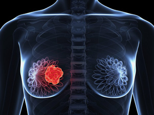
 Breast cancer is a kind of cancer that develops from breast cells. Breast cancer usually starts off in the inner lining of milk ducts or the lobules that supply them with milk. A malignant tumor can spread to other parts of the body. A breast cancer that started off in the lobules is known aslobular carcinoma, while one that developed from the ducts is calledductal carcinoma. Breast cancer is a kind of cancer that develops from breast cells. Breast cancer usually starts off in the inner lining of milk ducts or the lobules that supply them with milk. A malignant tumor can spread to other parts of the body. A breast cancer that started off in the lobules is known aslobular carcinoma, while one that developed from the ducts is calledductal carcinoma.The vast majority of breast cancer cases occur in females. This article focuses on breast cancer in women. Click here to read about breast cancer in men (male breast cancer). Breast cancer is the most common invasive cancer in females worldwide. It accounts for 16% of all female cancers and 22.9% of invasive cancers in women. 18.2% of all cancer deaths worldwide, including both males and females, are from breast cancer. Breast cancer rates are much higher in developed nations compared to developing ones. There are several reasons for this, with possibly life-expectancy being one of the key factors - breast cancer is more common in elderly women; women in the richest countries live much longer than those in the poorest nations. The different lifestyles and eating habits of females in rich and poor countries are also contributory factors, experts believe. According to the National Cancer Institute, 232,340 female breast cancers and 2,240 male breast cancers are reported in the USA each year, as well as about 39,620 deaths caused by the disease. |
| Diagnosing breast cancer |
Women are usually diagnosed with breast cancer after a routine breast cancer screening, or after detecting certain signs and symptoms and seeing their doctor about them. Below are examples of diagnostic tests and procedures for breast cancer:
Breast cancer screening has become a controversial subject over the last few years. Experts, professional bodies, and patient groups cannot currently agree on when mammography screening should start and how often it should occur. Some say routine screening should start when the woman is 40 years old, others insist on 50 as the best age, and a few believe that only high-risk groups should have routine screening. |
| Liver Cancer |
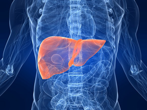
| Liver cancer is a condition that happens when normal cells in the liver become abnormal and grow out of control into cancer. Malignant or cancerous cells that arise out of liver cells are called hepatocellular carcinoma, and cancer that arises in the ducts of the liver is called cholangiocarcinoma. |
| How is liver cancer diagnosed? |
|
| HOME I ABOUT DR. I SERVICES I FAQ I CONTACT ME I ENQUIRY I DISCLAIMER |
| Site Powered By : www.calcuttayellowpages.com |

 The symptoms of mouth, head and neck cancers depend on where the tumour is found. Some common symptoms include:
 The best way to detect liver cancer is through surveillance ultrasound of the liver done every six months in a patient with a diagnosis of cirrhosis and to treat the liver cancer as soon as it is detected.