
A person with a ventral hernia has a piece of intestine that protrudes through an abnormal opening in the abdominal wall. In order for an ventral hernia to form, there must be a hole, tear or weakened area in the abdominal wall, usually caused by a previous surgical incision. This abnormal weakness allows the intestine to protrude through the hole or tear, and the intestine forms a bulge underneath the skin. The abnormal opening may be in the umbilicus or in a surgical scar. Causes of ventral hernia include brith defects, abdominal injuries, and abdominal surgery. Ventral hernias account for less than 5 percent of all hernias.
The most common symptom of a ventral hernia includes a bulge or knot under the skin in the abdomen. The bulge is more prominent while lifting, laughing, straining, or coughing. Usually, the bulge can be pushed back into the abdomen. Symptoms of a serious ventral hernia may include hernia pain or swelling, abdominal pain, abdominal swelling, vomiting, constipation, or fever.
A ventral hernia is treated with surgery, which may be performed with a laparoscope, or through an incision in the abdomen.
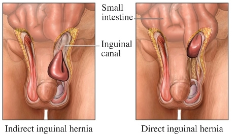
A hernia occurs when the inside layers of the abdominal wall weaken then bulge or tear. The inner lining of the abdomen pushes through the weakened area to form a balloon-like sac. This, in turn, can cause a loop of intestine or abdominal tissue to slip into the sac, causing severe pain and other potentially serious health problems.
Men and women of all ages can have hernias. Hernias usually occur either because of a natural weakness in the abdominal wall or from excessive strain on the abdominal wall such as strain from heavy lifting, substantial weight gain, persistent coughing, or difficulty with bowel movements or urination. Eighty percent of all hernias are located near the groin. Hernias might also be found below the groin (femoral), through the navel (umbilical), and along a previous incision (incisional).
A noticeable protrusion in the groin area or in the abdomen
Feeling pain while lifting
A dull aching sensation
A vague feeling of fullness
Nausea and constipation
Laparoscopic surgery uses a thin, telescope-like instrument known as an endoscope that is inserted through a small incision at the umbilicus (belly button). Usually, this procedure is performed under general anesthesia. This requires an evaluation of your general state of health, including a history and physical exam, possibly including lab work and EKG.
You will not feel pain during this surgery. The endoscope is connected to a tiny video camera, smaller than a dime, which projects an "inside view" of the patient's body onto television screens in the operating room. The abdomen is inflated with a harmless gas (carbon dioxide) to allow your doctor to view your internal structures. The peritoneum (the inner lining of your abdomen) is cut to expose the weakness in the abdominal wall. A mesh patch is attached to secure the weak area under the peritoneum. The peritoneum is then stapled or sutured closed. Following the procedure, the small abdominal incisions are closed with a stitch or two, or with surgical tape. Within a few months, the incision are barely visible.
Three tiny scars rather than one large abdominal incision
Short hospital stay (You might leave the day of surgery or the first day after surgery)
Reduced post-operative pain
Low hospital costs
Faster return to work
Shorter recovery time and earlier resumption of daily activities (a recovery time of days instead of weeks)
It is important to follow your doctor's instructions after surgery. Many people feel better in just a few days. However, you might need to take it easy for a week or two.
This procedure is as safe as open surgery, in carefully selected cases, when performed by specialists in this field.
